To install StudyMoose App tap and then “Add to Home Screen”
Save to my list
Remove from my list
This experiment was carried out to perform staining methods on microbes, explain the mechanisms of staining, namely, simple staining, and learn how to use the microscope. The experiment setting was kept as sterile as possible when conducting the experiment. The microbes under the microscope were drawn out and labeled. In conclusion, different staining techniques are used to determine the presence of certain exterior structures of the bacteria like the cell envelope or just to identify the basic shape and size of the microbe.
In this experiment, the microbes E. coli, Bacillus sp and an unknown microbe from petri dish 1 were categorized as gram negative or gram positive and their shape was identified. These staining techniques are the correct way to identify the shape and size of microbes.
Microbiology is the branch of biology that deals with microorganisms and their effect on other living organisms. Microbes are very small organisms which can only be viewed with the aid of microscope. Several groups of organisms that fit into this category are bacteria, cyanobacteria, fungi and protists.

Proficient in: Biology
 5 (876)
“ Have been using her for a while and please believe when I tell you, she never fail. Thanks Writer Lyla you are indeed awesome ”


Within this group there are several species interesting to humans because of their ability to cause disease or their use in the food industry and microorganisms can be classified to unicellular and multicellular. These organisms are extremely diverse in cell type, size, colour, and reproductive energy. Microbes can be classified by their cell type. All cells can be categorized either prokaryotic or eukaryotic and the primary difference between these two cell types is the presence of a membrane-bound nucleus.
The experiment was carried out to use a bright field microscopy, prepare and observe bacterial slides and perform staining methods and explain the mechanism of the bacteria.
In order to observe and investigate microbes we need to use microscope and bacterial Staining Techniques. Microscope is the invaluable tool allows the viewing of objects or structures that otherwise would go unnoticed by human naked eyes. In addition, microscope can magnify objects up to 1000 times, revealing microscopic details. It has special techniques and optics thus it can reveal the structure and biochemistry of living cells. Microscope consists of a combination of several optical lenses. In this experiment, we are using light microscope.
Light is conducted through curve lenses in such a way that an object may be viewed larger than its actual size. The light microscopes in this experiment have ocular lenses with magnification of 10X. Moreover, there are also four different objective lenses to choose from 10X, 40X, and 100X. For Bacterial Staining Method, there are two basics techniques. One of them is using wet mount method but bacteria are too small and too transparent to be well described using light microscopy and a wet amount. Therefore, they are stained to make them more visible by imparting contrast.
Simple stain, with only one layer of cell, is coloured with different colour and methylene blue dye are used for differentiating Bacillus sp. Negative stain is particularly useful for determining cell size and arrangement and it can be used to stain cells that are too delicate to be heat-fixed. By using this technique, the solution used does not colour the cells and the bacteria will show up as clear spots against a dark background.
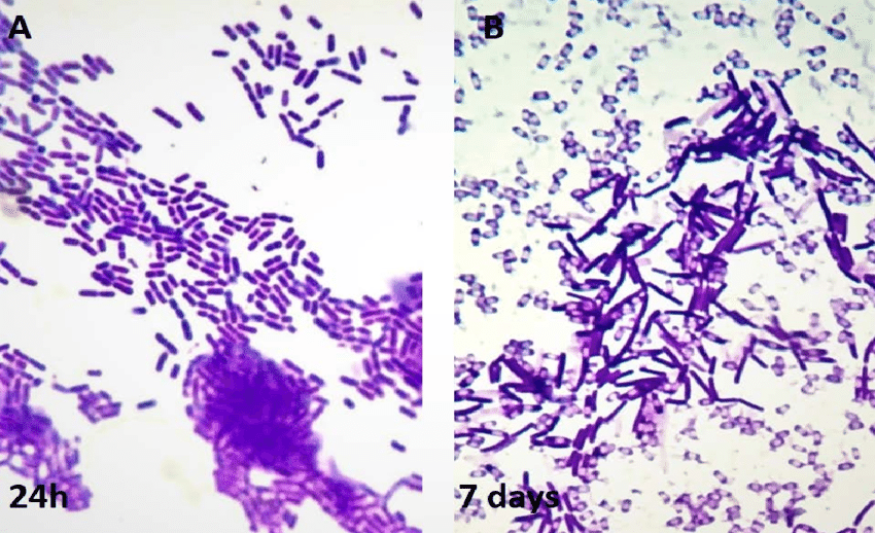
The Gram Staining Method is used as a tool for differentiation of Gram-positive and Gram-negative bacteria, as a first step to determine the identity of a particular bacterial sample. Gram-positive and Gram-negative organisms are distinguished from each other by differences in their cell walls, including the way the cell takes up and retains stains. The E. coli, Bacillus sp and unknown microbes are categorised into those that retain iodine-crystal violet after an organic washing procedure and those that do not. Gram Staining is the most consistent when done on bacteria that less than 24 hours, while the older cultures may not retain the primary stain and give inaccurate results. 2.0 Literature Review
Instrument that is used to see objects that are too small for the naked eyes is called microscope. It can magnify objects up to 1,000 times, revealing microscopic details. With special techniques and optics the structures and biochemistry of living cells can be revealed. There is various type of microscope, the most common and first to be invented is the optical microscope which uses light to image the sample. Although, Zaccharias Janssen discovered that object appeared greatly enlarged after experimenting with several lenses in a tube in 1590 and in 1609, Galileo worked out the principles of lenses, but Anton van Leeuwenhoek is a microscope designer who first to detect microorganisms using microscope. Light microscope employs visible light to detect small objects. The biggest challenge when it comes to looking at living things are obtaining sufficient contrast, finding the focal plane, obtaining good resolution and recognizing the subjects when one sees it.
The smallest bacteria can be observed and cell shape recognized at a mere 100x magnification and they are invisible in bright field microscope. Ocular lens is a cylinder containing two or more lenses. The function is to bring the image into focus for the eye. Staining is a process in which microbes are stained to enhance contrast in the microscopic image. Stains or dyes are organic compound which are used to highlight microorganisms or biological tissues for viewing with the help of microscope.
Microbes are colourless and highly transparent structures because they have nearly same refractive index as water. Therefore, microbes cannot be seen with our naked eyes thus different types of staining methods are used to increased visibility and contrast, accentuate specific morphological features, to detect extracellular and intracellular components of microbes and preserve them for future use. The basic requirements for staining are clean grease-free slide, bacteria tobe stained, inoculating loops and Bunsen burner to sterilise inoculating loops before and after smear preparation. Two ways to fixing the slides are heat fixation and chemical fixation. Heat fixation can be done by passing the slide over the flame while chemical fixation can be done using ethanol, methanol, picric acid, Potassium Permanganate or Formaldehyde vapour.
The function of fixation is to kills bacteria rendering safe handling and prevents autolysis by inactivating the autolytic enzymes. In addition, it increases the permeability of cells to stain, makes cell rigid and unfolds the globular proteins and exposing reactive groups and increasing affinity for stain. Different stains have different affinities for different organisms and they are used to differentiate different types of organisms. Bacteria are slightly negatively charged at pH 7.0 and basic dye stains bacteria while acidic dye stains background. For simple staining, only one dye is used. Simple staining is easier to perform but it has limitations. It was an easy method because only single staining agent used and either using basic or acid dyes. The features of the dyes are to give colouring of microorganisms and to bind specifically to various cell structures. For simple staining basic dyes which are positively charged are used. These dyes will attach to negatively charged
cytoplasm of microbial organism.
Figure 1 :
Bascillus sp
![]() Negative staining is particularly useful for determining cell size and arrangement. In addition, it can be used to stains cells that are too delicate to be heat-fixed. Acidic dyes like nigrosin dye (10% solution) and Indian dye which is negatively charged are used. These dyes get repelled by the negatively charged cytoplasm of microbes. Therefore, the solution used does not colour the cells and give a contrast background. It is commonly used for determining bacteria with capsules.
Negative staining is particularly useful for determining cell size and arrangement. In addition, it can be used to stains cells that are too delicate to be heat-fixed. Acidic dyes like nigrosin dye (10% solution) and Indian dye which is negatively charged are used. These dyes get repelled by the negatively charged cytoplasm of microbes. Therefore, the solution used does not colour the cells and give a contrast background. It is commonly used for determining bacteria with capsules.
Figure 2:
E. coli
Gram stain techniques identify bacteria as gram-positive which is the stain is retained or gram-negative which means the stain is washed. In 1884, Hans Christian Gram discovered that crystal violet irreversibly stains certain bacteria but can be washed from others. Gram staining can be described as a staining technique used to classify bacteria which is bacteria that are stained with crystal violet followed by a brief treatment with Gram’s iodine and after being decolourised with alcohol and treated with safranin then washed with water.
Those that retain the crystal violet are Gram-positive and those that do not retain it are Gram-negative. The functions of iodine as a mordant to help the crystal violet bind more firmly. Gram positive bacteria have the multiple layers of peptidoglycan retain the crystal violet while it is quickly rinsed out of Gram-negative bacteria because their peptidoglycan is a single layer thick. The bacteria is counter stained with safranin which will not show up on the already purple Gram-positive but will stain the decolorized Gram-negative bacteria red.
Figure 3: Cell wall difference
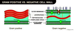
Cell wall difference
Microscope is turned on, and then the light source is adjusted. The objective lens was lowered till the lower far without touching the slide. By using the fastening clip the slide was fastened on the microscope stage. While looking at the eyepiece, the illuminator and diaphragm were adjusted. The coarse adjustment was slowly adjusted until the image of desire was focused. The slide was moved around to the centre of the field to focus to desire image. The slide is observed through the low power objective (x10), high dry objective (x40) and immersion oil objective (x100) for clearer view of culture. The stage is lowered to its minimum position after finished using the microscope. The switch is turned off and the microscope is covered back. B) Bacterial staining techniques
Alcohol soaked slide are run on the flame of Bunsen burner. One drop of water was dropped to the clean slide by using sterile loop. The cultures of Bacillus sp. are spread onto the water slide surface, again by using sterile loop. The loops were sterile again to kill excess microbe. The slide contained by culture was passed quickly through the flame of Bunsen burner for two second for each two or three time. The slide was flooded with methylene blue stain for a minute. The slide is rinsed with water and blotted to dry using bibulous paper. The prepared slide was examined under microscope by low power objective lens (x10), high dry power objective (x40) and oil immersion power objective (x100) to get the best view of the microorganisms. The morphology of the microorganisms was drawn.
The procedures were repeated with bacteria E. coli.
Alcohol soaked slide were run on the flame of Bunsen burner. The Bacillus sp. cultures are spread onto the slide surface, by using sterile loop. The slide was set to air dry without exposed to heat. Two to three of Nitrogen dye solutions were dropped to the smear. The Nitrogen dye solutions were spread by using the edge of another slide side became one thin film. The prepared slide was let for air dry.
The prepared slide was examined under microscope by low power objective lens (x10), high dry power objective (x40) and oil immersion power objective (x100) to get the best view of the microorganisms. The morphology of the microorganisms was drawn.
The procedures were repeated with bacteria E. coli.
Alcohol soaked slide were run on the flame of Bunsen burner. The Bacillus sp. cultures were spread onto the slide surface, by using sterile loop. The slide was set to air dry and exposed to heat for few seconds. The slide was added with crystal violet solution and let for one minute. The slide was rinsed with water and flooded with iodine solution for one minute. The slide was washed with water and added with decolourizer till crystal violet colour disappeared. The slide the rinsed with water. Safranin was added and waits for a minute. Then the prepared slide was washed with water for maximum 5 seconds. The prepared slide was dried with bibulous paper and allowed to air dry. The prepared slide was examined under microscope by low power objective lens (x10), high dry power objective (x40) and oil immersion power objective (x100) to get the best view of the microorganisms. The morphology of the microorganisms was drawn.
The procedures were repeated with bacteria E. coli. and sample E2.
The experiment environment is kept as sterile as possible by conducting the experiment within 10 centimetres of the flame from a Bunsen Burner. This is to avoid contamination of the sample by microbes in the air. Gloves are worn for the same purpose. The loops used to smear the microbes onto the slide are sterilised three times in a flame. Meanwhile, the slid with sample on was to fix the bacteria onto the slide. Now about the stains, methylene blue dye is used to make the cells and nuclei more visible6. Crystal violet is used to stain the cell walls of bacteria6, which consist of peptidoglycan.
Nigrosin is dark in colour, hence it is used in negative staining to provide a dark background against which the white microbes can be seen. In Gram staining, cells are stained with crystal violet dye. Next, a Gram's iodine solution (iodine and potassium iodide) is added to form a complex between the crystal violet and iodine. This complex is a larger molecule than the original crystal violet stain and iodine and is insoluble in water. Alcohol is added to the sample as a decolouriser, which dehydrates the peptidoglycan layer, shrinking and tightening it. The large crystal violet-iodine complex is not able to penetrate this tightened peptidoglycan layer, and is thus trapped in the cell in Gram positive bacteria.
Conversely, the outer membrane of Gram negative bacteria is degraded and the thinner peptidoglycan layer of Gram negative cells is unable to retain the crystal violet-iodine complex and the colour is lost. A counterstain, safranin, is added to the sample, staining it red. Since the safranin is lighter than crystal violet, it does not disrupt the purple coloration in Gram positive cells. However, the decolorized Gram negative cells are stained red. Gram positive bacteria (with a thicker peptidoglycan layer) retain crystal violet stain during the decolourisation process, while Gram negative bacteria lose the crystal violet stain and are instead stained by the safranin in the final staining process.6 From our observation from simple staining of Bacillus sp ,it can be seen that the bacteria is rod-shaped.
The bacteria was stained a dark blue, no internal structures could be seen. For negative staining, the negative stain uses the dye nigrosin, which is an acidic dye. By giving up a proton (as an acid) the chromophore of the dye becomes negatively charged. Because the cell wall is also negatively charged only the background around the cells will become stained, leaving the cells unstained. Hence, the cells are seen as white spots against a dark background E.coli is a rod shape as well. After gram staining for E.coli, E.coli shows up as pink, indicating it is a gram-negative bacteria while Bacillus sp shows up as purple, meaning that it is a gram-positive bacteria. The unknown microbes from petri dish
1turn out to be a mixture of gram negative and gram positive bacteria because of the presence of light pink and purple regions. They are round in shape.
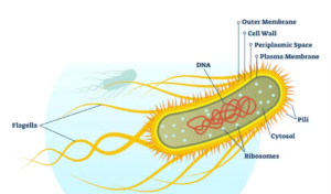
Morphology includes size, cell structure, presence of endospore and flagella. From the report, in Figure 8, the unknown microbes from petri dish 1 give purple and pink colouration after gram staining, they’re round in shape. A more reliable method to identify cell morphology would be to use special stains to identify specific parts of a microbe like endospores, which is usually present in gram positive bacteria. The method use to stain endospores is called the Schaeffer-Fulton1 method, where Malachite Green is used to stain the endospores while Safranin is a counterstain. The end result would be pink bacteria with green dots within them. Three methods to characterise a microorganism include:
This bio-chemical test is used on gram-positive bacteria to identify bacteria that can hydrolyze starch (amylose and amylopectin) using the enzymes a-amylase and oligo-1,6-glucosidase. Often used to differentiate species from the general Clostridium and Bacillus. Because of the large size of amylose and amylopectin molecules, these organisms cannot pass through the bacterial cell wall. In order to use these starches as a carbon source, bacteria must secrete a-amylase and oligo-1,6-glucosidase into the extracellular space.
These enzymes break the starch molecules into smaller glucose subunits which can then enter directly into the glycolytic pathway. In order to interpret the results of the starch hydrolysis test, iodine must be added to the agar. The iodine reacts with the starch to form a dark brown colour. Thus, hydrolysis of the starch will create a clear zone around the bacterial growth. E.g. Bacillus subtilis is positive for starch hydrolysis.2 ii) Protein analysis (gel electrophoresis, SDS-PAGE, establishment of clonality) The size and other differences between proteins among different organisms can be determined by using protein separation methods, collectively known as gel electrophoresis.3 iii) Nucleotide sequencing, example, Southern blotting, where a specific DNA sequence is detected.4 3)
Full standard procedure for operating a microscope:
When moving a microscope, always carry it with both hands. Grasp the arm with one hand and place the other hand under the base for support. ii. Turn the revolving nosepiece so that the lowest power objective lens is "clicked" into position. iii. The microscope slide should be prepared with a coverslip or cover glass over the specimen. This will help protect the objective lenses if they touch the slide.
Place the microscope slide on the stage and fasten it with the stage clips. iv. Look at the objective lens and the stage from the side and turn the coarse focus knob so that the objective lens moves downward (or the stage, if it moves, goes upward). Move it as far as it will go without touching the slide. v. Look through the eyepiece and adjust the illuminator (or mirror) and diaphragm for the greatest amount of light. vi. Slowly turn the coarse adjustment so that the objective lens goes up (away from the slide).
Continue until the image comes into focus. Use the fine adjustment, if available, for fine focusing. If the microscope has a moving stage, then turn the coarse knob so the stage moves downward or away from the objective lens. vii. Move the microscope slide around so that the image is in the center of the field of view and readjust the mirror, illuminator or diaphragm for the clearest image. viii. Then change to the next objective lens with only minimal use of the focusing adjustment. Use the fine adjustment, if available. If you cannot focus on your specimen, repeat steps 4 through 7 with the higher power objective lens in place. ix.
The proper way to use a monocular microscope is to look through the eyepiece with one eye and keep the other eye open (this helps avoid eye strain). x. Do not touch the glass part of the lenses with your fingers. Use only special lens paper to clean the lenses. xi. When finished, lower the stage, click the low power lens into position and remove the slide. xii. Always keep your microscope covered when not in use. Dust is bad for the microscope. 4) Three accidents that can occur during the experiment are: i. A sleeve catching fire while passing the slide through the flame in wide sweeping motions ii. the slide dropping due to a weak grip on it
The slide breaking due to over-exposure to fire and a strong grip.
The staining techniques in this experiment are the correct way to identify the shape and size of the bacteria. Bacillus sp is rod-shaped and gram positive while E.coli is rod-shaped and gram negative. The unknown microbes from petri dish 1 are a mixture of gram-positive and gram-negative bacteria and are round.
It is suggested that a smaller amount of microbes are smeared on the glass slide to prevent the sample from looking so dense under the microscope, thus preventing us from seeing the shape and size clearly. Next, increase the amount of the negative stain to ensure more visibility of the cells under the microscope. Lastly, clean the lens of the microscope before use to avoid confusing images in the eyepiece.
Lab Report about Simple Staining of Microbes. (2024, Jan 10). Retrieved from https://studymoose.com/document/lab-report-about-simple-staining-of-microbes
👋 Hi! I’m your smart assistant Amy!
Don’t know where to start? Type your requirements and I’ll connect you to an academic expert within 3 minutes.
get help with your assignment

