StudyMoose App
24/7 writing help on your phone
Save to my list
Remove from my list
This assignment is about the macromolecules and biological principles, it divides into three parts, firstly, it will identify the sickle cell anaemia disease, analyse and explain its cause. Afterwards, it will explain the Inheritance pattern of the condition following Mendel’s laws and the treatments available for the sickle cell patients. The second part is about the cell membrane, it outlines the chemical structure of Phospholipids, protein and carbohydrates, and its role in membrane functioning. The essay will develop into Aerobic Respiration in the last sections, by describing the structure of enzymes relating to their function.
It will then outline the key stages of aerobic respiration and explain how AdenosineTriphosphate (ATP) is produced. In conclusion, the main points are linked together and summarise the founding.
Description generated with very high confidenceSickle Cell Disease (SCD) is a group of disorders that affects haemoglobin (figure 1), the molecule in red blood cells that contain normal haemoglobin are disc shaped, and it transports oxygen to cells throughout the body.

 5 (234)
“ Very organized ,I enjoyed and Loved every bit of our professional interaction ”


The shape allows the cells to move through large and small blood vessels to deliver oxygen ( NHS, 2016). Haemoglobin can form stiff rods within the red cell. They are not flexible and stick to vessel walls, inflicting a blockage that slows the flow of blood and oxygen (figure 2 and 2a) (NHS, 2016).
The base changes that take place and convert round blood to sickle cell blood takes the form of the code in the molecules of DNA (deoxyribonucleic acid) and copied into one form of RNA. (ribonucleic acid) then to the final form to make protein which build the cell and control its action.
(Fullick 2011) as shown on figure 3)
DNA is the hereditary material responsible for passing genetic information from cell to cell, and generation to generation. Also it provides the information inherited by daughter cell offspring. That require the DNA to serve as a template for the transcription of the information into RNA and for the replication of the information into daughter DNA molecules. (Michigan University 2013) DNA is double stranded structures, which are complementary therefore, adenine (A) always pairs with thymine (T) or uracil (U) in RNA, and guanine (G) pairs with cytosine (C) (Meisenbergetal,2012) RNA is a polymer made up of nucleotide. It is a single, short polynucleotide chain where the pentose sugar is ribose and the base are adenine, guanine, cytosine and uracil. It has two types which are important in protein synthesise. Messenger RNA (mRNA) and transfer RNA (tRNA) (figure 4) (Toole 2015)
Due to single nucleotide substitution (adenine to thymine) in the codon of amino acids 6 of globin; this change (figure 5) converts a glutamic acid codon (GAG) to a valine codon (GTG) this form of haemoglobin is referred to as HbS, and normal adult haemoglobin is referred to as HbA. Substitution of a hydrophobic (valine) for a polar residue (glutamic acid) results in deformation of the red blood cell into a sickle-like shape making it relatively inflexible and unable to easily traverse the capillary beds. (Fry 2010)
The underlying problem in SCD is that the valine for glutamic acid substitution results in haemoglobintetramers that aggregate into arrangements upon deoxygenation in the membranes. Repeated cycles of oxygenation and deoxygenation lead to irreversible sickling (The medical biochemistry, 2017).
The absence of a polar amino acid promotes the non-covalent aggregation of haemoglobin in a low-oxygen environment which decreases their elasticity, which causes a monogenetic disorder that is created by a mutation in a single gene. The mutation could be present on one or both chromosomes (one chromosome inherited from each parent) (Genetic home, 2017).
Signs and symptoms of sickle cell disease more often start at a young age. Characteristic of this disorder include a low number of red blood cells, repeated infections, and intermittent periods of pain. Few people have mild symptoms, while others are frequently hospitalised for more severe complications (Ballas, 2015).
The signs and manifestation of symptoms are when red blood cells sickle, they separate prematurely, which can prompt to anaemia. Anaemia can cause shortness of breath, weariness, and delayed growth and development in children (Genetics Home Reference 2017).
Description generated with high confidenceSCD can be inherited from Autosomal Recessive Pattern (ARP) (figure 6), which means: one of few different ways that a trait, disorder, or disease can be passed down through families (Medlineplus, 2018). The parents of an individual with an ARP each carry one copy of the mutated gene, but they typically do not manifest signs and symptoms of the condition. In the ARP There is a 25% chance that one of the children could be born with SCD.
There is also 25% of an unaffected child, and 50% chance of one child will get the sickle cell trait (figure 7). That follows Mendel's law of inheritance patterns in pea plants that can be used to determine whether the disease-associated gene is located on an autosome or a sex chromosome (Nature, 2008). Mendel discovered the principal laws of inheritance, he derived that genes come in pairs and are inherited as distinct units, one from each parent (Omim, 2010). Mendel followed the segregation of parental genes and their appearance in the offspring as dominant or recessive traits(Macy, 2018). Mendel's Law of Heredity a gene pair defines each inherited trait, parental genes are randomly isolated to the sex cells, so that sex cells contain just a single gene of the pair (Medicine, 2016). Offspring, therefore, inherit one hereditary allele from each parent when sex cells unite in fertilisation. This process is known as meiosis (UpToDate 2016).
Carrier and normal, where one parent has sickle cell trait (HbAS), and the other does not carry the sickle haemoglobin at all (HbAA) There is (50%) chance one child will have one copy of the (HbAS) gene and have the sickle cell trait (King, 2017) ( figure 7) .
Carrier and Sickle Cell Anaemia: If one parent is (HbAS) carrier and the other has sickle cell anaemia (HbSS) there is 50% chance that one child will get sickle cell trait and one child will get sickle cell anaemia. No children will be completely healthy (figure 8) (Sickle cell society, 2014).
The treatments of the sickle cell include antibiotics, painkillers, routine vaccinations and blood transfusions. A new treatment, hydroxyurea, produces fatal haemoglobin, which helps prevent red blood cells of sickling. At the moment, the only cure for the disease is Bone Marrow Transplantation; where the affected person is transplanted with bone marrow from their healthy, genetic matching sibling. However, the procedure can cause complications (National Human Genome Research Institute, 2016).
The cell membrane, also known as the plasma membrane or cytoplasm membrane, is a double layer of lipids, proteins and carbohydrates that surrounds all living cells, arranged in a fluid mosaic structure, as shown in (Murphy et all, 2011) (figure 10). It is selectively permeable, which means that it only lets specific molecules a chance to enter and exit. It can likewise control the amount of some substances that go into or out of the cell (biology corner, 2004).
The cell membrane is a barrier that isolates a cell from its surrounding environment. It is made of four different types of molecules:
The fluid mosaic model portrays the structure of a cell membrane. It demonstrates that the cell membrane is not solid. It is flexible, and it moves… The plasma membrane is a mosaic of phospholipids, cholesterol molecules, proteins and carbohydrates (Bownessetal, 2008)
Phospholipids consist of two hydrophilic polar heads group and two hydrophobic tails. The phosphate heads contain one or more phosphate groups (PO43-) (figure 11). The hydrophobic tails are made up of two fatty acid chains. When numerous phospholipid are placed in water, their heads tend to face the water, and the tails are compelled to stick together, forming a bilayer, organised in a manner that keeps them away from water. The components are connected via third molecule, glycerol. (figure 13) (Fullick, 2012)
Functions of Phospholipids in cell membrane; Phospholipids procure barriers to protect and secure the cell, and they make barriers for the organelles inside those cells. Phospholipids work to give pathways for various substances across membrane, meaning that only certain molecules can pass through it to enter or exit the cell.
Phospholipids maintain a gradient of chemical and electrical processes to ensure cell survival (Murphy et all, 2011). In the human body, phospholipids are found in areas like the lung and in joint where it helps to lubricate the cells. It is used a delivery system which help to transport drugs throughout the body to the area which is in pain. In animals, phospholipid is present in all mammalian tissues and in all parts of the cell, including mitochondria, microsomes and nucleus. The bacteria membrane is represented by the 40% of phospholipids, and they have their own membrane. A plant hormone is similar in structure to phospholipid, which are found in high concentration such as soybeans, sunflowers, cotton seed, corn … (Sinclair, 1938)
The cell membrane contains two types of related proteins. Peripheral proteins are exterior and associated to the membrane by connection and interactions with other proteins. The Integral ones are inserted into the membrane, and most go through it. Cell membrane proteins have various different functions. Their main functions are to give the cell support and shape. They help the cells to communicate with their external environment (figure 13).
Proteins they are a most abundant molecule in the cell. They have four structures level; primary structure; a sequence of amino acids in the polypeptide bond. Secondary structure; the polypeptide bonds do not remain flat. The tertiary structure; the amino acid coiled and foiled. Quaternary structure; is where different polypeptide chains hold together by bonds. (figure 14) ( Nature Education, 2014)
Function of proteins in cell membrane; Proteins comprise about 50% of the mass, They involved in transporting substances across the membrane. They are maintaining the cell's shape, or in cell motility. They may also be enzymes catalysing reactions in the cytoplasm. They also can act as receptors by having a specific binding site where hormones or other chemicals can bind (Sparknote, 2017).
The proteins is found in human, plant and bacteria; in the human the proteins are composed by 16% of the cells, they have many critical roles and crucial to good health, There are 20 amino acids that provide thousands of different proteins in human body. Proteins do most of their work in the cell and perform various jobs such as acting as messenger, causing biochemical reactions and the growth and maintenance of a cell (Meisenberg & Simmons, 2012).
Plants store proteins to provide carbon, nitrogen, and resources for subsequent growth and development, the amino acids that composed the protein are crucial for a plant live cycle. In a bacterial cell the protein really important and most of them are found in the cytoplasm, ribosomes, flagella and cell wall… They have quite the same role as in the human body.
The carbohydrates (figure 14) are consist of the elements carbon (C), hydrogen (H) and oxygen (O) with a proportion of hydrogen twice that of carbon and oxygen. Carbohydrates is the main sources of ATP and has numerous other compounds found in living organisms. In their main form, carbohydrates are simple sugars or monosaccharides that are metabolized by certain enzymes. The combination of two simple sugars is a disaccharide. Carbohydrates including of two to ten simple sugars are called oligosaccharides, and those with a more significant number are called polysaccharides (Scientific psychic, 2017).
Functions of carbohydrate perform two main functions: participate in cell recognition and adhesion, either cell signalling or cell-pathogen interactions, and they have a structural role as a physical barrier. Starch is the primary energy storage material in the plant. Glycogen is the primary energy storage material in animals (Science direct, 2012). The glycocalyx is involved in protection and cell recognition, and antigens such as the ABO antigens on blood cells. Glycoproteins play role in the recongnition of hormones and foregin molecules. Strach is the main energy storage material in plant. Glycogen is the main energy storage material in animals (science direct 2012).
In plants cell they have two main direction; they are building blocks for plants structural components per examples cellulose; they help to give energy to the plant cell… In human body the carbohydrates are critical to support life’s most basic function and the primary body source of fuel, by providing energy production, its storage and It helps to build macromolecules.
The key stages of aerobic respiration and ATP production
Enzymes are large molecules, made of large chains of amino acids which have been folded into specific shape. Their function is related to their structure. A substrate is a molecule where an enzyme acts; they fit perfectly together (Worthing Biochemistry, 2017). The surface which the substrate aligns itself is known as the active site. The enzyme can have no protein group which is known as a cofactor. Its present is essential for the enzyme to function. Apoenzyme is the protein component of an enzyme separable from the cofactor but requires its presence to form holoenzyme, which is complete and catalytically active. The lock and key, the action of enzymes come up with the lock and fundamental model where the substrate fits in enzyme-like key and lock. The induce fit, it makes the active site change the shape in the right way so the substrate can fit in. (figure 14) (Philips,2016)
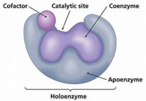
Figure 15 shows the chemical structure of enzyme
Enzymes are proteins that take part in cellular metabolic processes with the ability to enhance the rate of reaction between bio-molecules. They lower the Activation energy by bringing specific molecules together rather than relying on random collision. (Chemguide, 2016) (figure 16)
PH also affect enzyme activity. However, some enzymes can work in extreme PH, e.g. Protease enzyme in animals. PH affects the charge of amino acids at the active site, so the substrate will no longer bind (figure 17) (City University of New York, 2016).
Figure 17 shows and explains how temperature affects the enzyme reaction.
Image result for ph in enzymes Figure 16 shows the affect of PH in anzyme reaction
Image result for temperature in enzymes
The aerobic respiration and ATP production
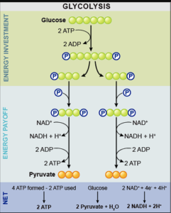
The metabolic pathway involved in respiration can be divided into three parts: Glycolysis (figure 18) it occurs in the cytoplasm. Is the breakdown of glucose into two pyruvate molecules, the process does not require oxygen it is (anaerobic) the production of pyruvate involves the production of several neutral molecules. Phosphorylation of some these intermediates requires 2 ATP molecules in an energy investment stage. 4 ATP molecules are regenerated in the production of other intermediates. The breakdown of glucose into pyruvate results in a net gain ATP molecule. Dehydrogenase enzymes remove electrons from the intermediates, and these electrons are transferred to the enzymes NAD forming NADH. Those co-enzymes take the electrons to the inner membrane of the mitochondria for use in the Electron Transportation Chain. If oxygen is available pyruvate molecules progress into the (TCA). If oxygen is not available the pyruvate undergoes fermentation, which can be Alcoholic such as ethanol and CO2 or Lactic Acid such as lactic acid ( biology innovation, 2008). the overall of glycolysis: Glucose + 2xADP + 2xNAD+ -> 2xPyruvate + 2xATP + 2xNADH (Glycolysis, 2011) .
Figure 18 shows the Glycolysis respiration pathway.
Citric Acid Cycle (tricarboxylic acid) (TCA) occurs in the matrix of mitochondria. It requires oxygen (aerobic) the pyruvate enters the matrix of the mitochondria and carbon dioxide is removed. Acetyl group are formed and combines with coenzyme A to form coenzyme A acetylene. Coenzyme A connect with a molecule called Oxaloacetate to form Citrate. The cell regenerates Oxaloacetate by breaking down citrate using an enzyme controlled series of reactions. Intermediate molecules are formed because the enzymes remove carbon and hydrogen electron, the carbon dioxide is released as a by-product the citric acid also results in the creation of ATP. Dehydrogenase enzymes remove hydrogen Ions and electrons from intermediates, which passed to coenzymesNAD or FAD forming NADH or FADH the high energy electrons are passed to the (ETC) The overall reaction of the pathway is: 2 acetyl CoA + 6NAD+ + 2FAD + 2ADP + 2H3PO4 4CO2 + 6 NADH + 6H+ + 2FADH2 + 2ATP as shown on (figure 18) (Royal Society of Chemistry, 2004).
Electron Transport Chain (ETC) is the last stage of the respiration pathway, and it produces the most ATP molecules. It occurs in the inner membrane of mitochondria. NDH, FADH2 release the electrons into the transport chain. The electrons transfer energy to the proteins of the membrane providing the energy for hydrogen ions to be pumped across the membrane. The flow of the ions across the membrane synthesises ATP by ATP synthase. 3 ATP is produced from each NADH, and 2 ATP are produced from each FADH which transfers high energy electrons to the ETC this results in a total of 34 ATP Molecules. The overall of the pathway is 10NADH + 2FADH2 34ATP. (figure 19) Total of 38 ATP is produced from the three stages of the respiration pathway. (Khan academy, 2011)
To conclude, due the single nucleotide change in the codon of amino acid 6 ( GAG- GTG) it result the deformation of red boold cell into sickle shape like, and it can be passed from the parents to their children in different inheritance pattern. The cells require many compounds to survive, these compounds are protein, carbohydrates and phospholipids, they are very important in every living organisms as much as the respiration.
Understanding Sickle Cell Anaemia: Genetics, Cell Membrane, and Aerobic Respiration. (2024, Feb 22). Retrieved from https://studymoose.com/document/understanding-sickle-cell-anaemia-genetics-cell-membrane-and-aerobic-respiration
👋 Hi! I’m your smart assistant Amy!
Don’t know where to start? Type your requirements and I’ll connect you to an academic expert within 3 minutes.
get help with your assignment
